Medically reviewed by a board-certified general surgeon. Educational content only; it does not replace an examination by your own physician.
You noticed a small, firm lump just above your belly button. It may ache after lifting, or only show itself when you cough or sit up from bed. Perhaps you were told it was “just a muscle strain” or “weight gain.” As a general surgeon, I see this exact story every week, and I want you to leave this article knowing what the lump is, why it formed, and what to do next.
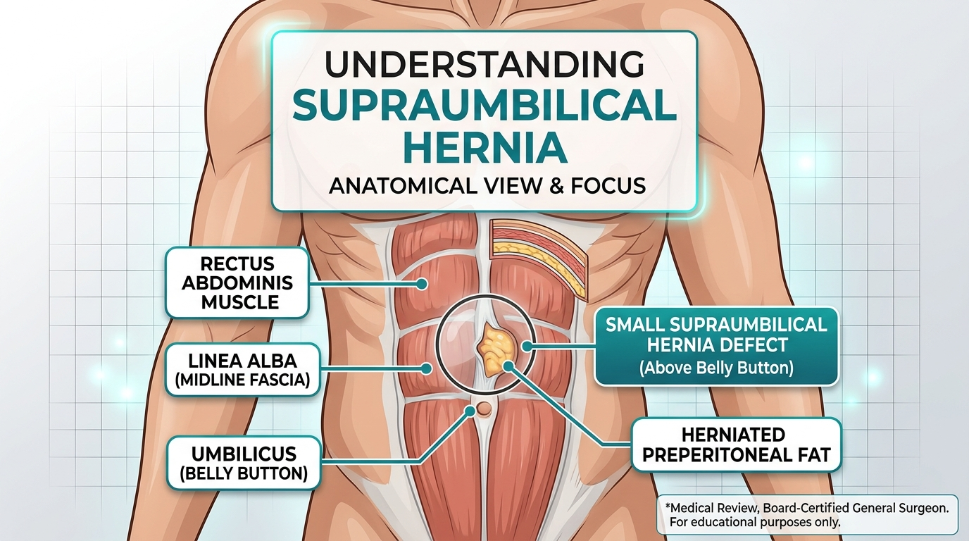
The short answer: a supraumbilical hernia forms when pressure inside the abdomen pushes fat, or occasionally bowel, through a weak point in the linea alba, the midline tendon sheet between the two rectus abdominis muscles, in the area just above the umbilicus. The weakness is usually a combination of a congenital gap, repeated strain, weight, pregnancy, and tissue quality. It rarely has a single cause.
Understanding the Condition: What Is a Supraumbilical Hernia?
The abdominal wall is a layered structure. In the midline, the two rectus abdominis muscles are joined by the linea alba, a fibrous band formed from the interwoven aponeuroses of the abdominal muscles. A hernia is a defect in that wall through which tissue protrudes.
- Location: the defect sits in the linea alba, just cranial to the umbilical ring.
- Classification: the European Hernia Society (EHS) classification counts a midline hernia within 3 cm above or below the umbilicus as an umbilical hernia, and one further up toward the xiphoid process as an epigastric hernia [1]. Supraumbilical hernias sit on the border between the two, which is why you will see different names for the same problem.
- Contents: most often preperitoneal fat or omentum. Less commonly, a loop of small bowel.
- Primary vs. incisional: a primary hernia appears in unoperated tissue. An incisional hernia forms at a prior surgical scar, such as a laparoscopic port site or a midline laparotomy. Their causes differ, and I cover both below.
What Causes a Supraumbilical Hernia in Adults?
Every abdominal wall hernia comes down to one equation: intra-abdominal pressure versus tissue strength. Either the pressure is too high, the wall is too weak, or both.
1. Structural weakness of the linea alba
The linea alba is not uniform. Anatomical and biomechanical studies show it is narrower and differently woven in places, and that small natural gaps for perforating vessels exist along its length [2]. In some people these gaps never close fully. You can carry a latent weak point for decades before a trigger exposes it.
2. Repeated or sudden spikes in intra-abdominal pressure
- Heavy lifting, especially with breath-holding (the Valsalva manoeuvre)
- Chronic cough from smoking, COPD, or asthma
- Chronic constipation with straining
- Obstructive urinary symptoms, such as an enlarged prostate
- Intensive core training with poor bracing technique
3. Pregnancy and diastasis recti
Pregnancy stretches the linea alba. Many women develop diastasis recti, a widening of the inter-rectus distance. Diastasis is not itself a hernia, but a thinned, stretched midline is a perfect site for one to appear years later. If you have both, the plan for repair changes (see the treatment section).
4. Obesity and abdominal fat
Visceral fat raises resting intra-abdominal pressure around the clock and thins the tissues over time. Preperitoneal fat is also the usual “content” of these hernias.
5. Smoking and tissue quality
Smoking impairs collagen synthesis and wound healing, and it is an established risk factor for hernia formation, particularly incisional hernias. Diabetes, corticosteroid use, ageing, and connective tissue disorders such as Ehlers-Danlos syndrome add to this.
6. Previous surgery
Laparoscopic port sites and midline incisions above the navel are classic sites for incisional hernias. Infection, poor closure technique, or post-operative straining all raise the risk.
7. Fluid and rapid weight change
Ascites (from liver disease) and large swings in weight stress the abdominal wall. Patients with ascites need specialist assessment before any elective repair.
How Does a Supraumbilical Hernia Develop? A Step-by-Step Guide
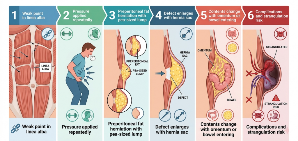
- A weak point exists. This is a congenital gap or a stretched area of the linea alba.
- Pressure is applied repeatedly. Coughing, lifting, straining, or weight gain pushes outward.
- Preperitoneal fat herniates first. A small plug of fat passes through the gap. This often causes a tender, pea-sized lump.
- The defect enlarges. Each pressure surge widens the gap, and the hernia sac, a pouch of peritoneum, may follow.
- Contents change. Omentum or bowel may enter the sac. At this point the risk of incarceration (stuck contents) rises.
- Complications can occur. If trapped tissue loses its blood supply, the hernia becomes strangulated, a surgical emergency.
Symptoms: What You Might Notice
- A lump above the navel that is more visible when standing, coughing, or tensing your abdominal muscles
- A dull ache or sharp “catching” pain at the lump, often worse late in the day
- A lump that you can push back in (reducible) or that no longer goes back in (irreducible)
- Tenderness over the lump, which with a small fatty hernia can be surprisingly intense
Clinical Note: How this hernia affects daily life. Small midline hernias are often dismissed because they are “only a little lump.” Yet I regularly hear that patients have stopped exercising, avoid bending to tie shoes, wear loose clothing to avoid pressure on the area, and sleep poorly because of a pulling sensation when turning in bed. Some patients lose appetite because a full stomach presses on a tender spot, and many become anxious about the lump itself. These effects are real, they are medically valid, and they are a legitimate reason to seek repair, even when the hernia is small.
Why It Is Often Misdiagnosed
Supraumbilical and epigastric hernias are frequently missed or mislabelled because the symptoms are vague and the lump can be tiny
- “Gastritis” or “reflux”: upper-midline pain after meals is blamed on the stomach.
- “Muscle strain”: the pain is attributed to a pulled abdominal muscle.
- “Lipoma”: a fatty hernia feels like a fatty lump under the skin.
- Normal ultrasound or CT scan: small defects can be missed if the scan is done lying down and without a cough or Valsalva manoeuvre.
- Diastasis recti: the midline bulge of diastasis can hide a true hernia inside it.
A dynamic ultrasound, performed standing with the patient straining, is a quick and accurate way to confirm a hernia when examination is uncertain.
From My Clinical Experience: A Patient’s Journey
The patient: a 41-year-old teacher and mother of two, with a body mass index of 29. For eight months she had burning pain above her navel after meals.
What she was told: she was treated for gastritis with a proton pump inhibitor, then told she had a pulled muscle from the gym. An abdominal ultrasound while lying still was reported as normal. By the time she reached me, she had stopped exercising and was avoiding family meals because of the pain.
What I found: when she stood and coughed, a firm, tender, 1.5 cm lump appeared 2 cm above the umbilicus. A standing dynamic ultrasound confirmed a linea alba defect of about 1.2 cm containing preperitoneal fat. She also had mild diastasis recti.
Treatment: we discussed the options. Because the defect was larger than 1 cm, I recommended a mesh repair. We performed a small open preperitoneal (sublay) mesh repair under general anaesthesia as a day case.
Outcome: she went home the same day, returned to desk work after one week, and to full activity at six weeks. At her three-month review the pain was gone and she was eating normally again. Her words: “I didn’t realise how much I’d been bracing against that pain every day.”
Treatment Options: Comparing the Repairs and Why I Choose Between Them
Watchful waiting
A very small, painless, easily reducible hernia can be monitored. The EHS and Americas Hernia Society (AHS) guidelines accept this approach in selected, asymptomatic patients, but you must know the warning signs below.
Open suture repair vs. open mesh repair
- Suture-only repair closes the defect with stitches. It is quick, but recurrence is higher. In a landmark randomised trial of midline incisional hernias, suture repair had a 3-year cumulative recurrence of 43% compared with 24% for mesh.
- Mesh repair reinforces the closure with a synthetic mesh. Current guidance recommends mesh for primary umbilical and epigastric defects of 1 cm or larger, and it is increasingly favoured for smaller ones as well.
My practice: for a defect under 1 cm in a slim, non-smoking patient, a careful suture repair is reasonable. For anything 1 cm or larger, a coexisting diastasis recti, obesity, or a recurrence, I use mesh. This is because the tissue around these defects is often weakened, and stitching weak tissue together under tension tends to fail.
Open vs. laparoscopic/robotic repair
| Factor | Open (sublay or onlay) | Laparoscopic / robotic |
| Incision | Single small midline incision | 3 small incisions |
| Best for | Small to medium defects, thin patients | Multiple defects, larger defects, diastasis, obese patients |
| Mesh placement | Preperitoneal (behind the muscle) | Intraperitoneal or preperitoneal |
| Trade-offs | Wound complications are somewhat more likely | Needs general anaesthesia; more expensive; small risk of bowel injury |
I favour open preperitoneal repair for most single, small supraumbilical hernias because it avoids contact between mesh and bowel and needs only one small incision. I move to a minimally invasive approach when there are several defects, when a diastasis needs correction, or when I want to reduce the risk of wound infection in a patient with obesity or diabetes.
Recovery Timeline
- Day 0: home the same day in most cases. Expect soreness and mild bruising.
- Days 1–7: walk several times a day. Take simple analgesia. Avoid lifting anything heavier than a kettle.
- Weeks 1–2: return to desk work. Keep the wound clean and dry.
- Weeks 2–4: gradually increase walking and light daily tasks.
- Weeks 4–6: most patients resume lifting and the gym, building up slowly.
- Month 3: the scar has settled. Full activity is expected.
Risks, Honesty, and Realistic Expectations
Every operation carries risk. Specific risks of hernia repair include:
- Seroma (a fluid collection) or haematoma
- Surgical site infection, which is higher in smokers and diabetics
- Chronic pain at the repair site, a small minority of patients
- Recurrence, which depends on defect size, mesh use, smoking, and body weight
- Mesh-related complications, which are uncommon
Stopping smoking at least 4 weeks before surgery and optimising weight and blood sugar are the most effective things you can do to improve your result.
When to Seek Emergency Care
Go to the emergency department immediately if your hernia:
- Becomes suddenly painful, hard, and cannot be pushed back
- Turns red, purple, or dark
- Occurs with vomiting, a swollen abdomen, or inability to pass wind or stool
- Occurs with fever
These are signs of incarceration or strangulation, which require urgent surgery. Do not wait to see if they settle.
Supraumbilical Hernia Causes: Common Questions Answered
Can a supraumbilical hernia go away on its own?
No. A fascial defect does not heal without repair. It can remain stable for years, but it will not close.
Is a supraumbilical hernia caused by sit-ups?
Sit-ups do not usually create the weakness, but they can reveal a pre-existing one by repeatedly raising intra-abdominal pressure.
Who is most at risk? Use cases and applications for risk assessment
- People who are overweight or obese
- Smokers
- People who have been pregnant, especially more than once
- Those with a chronic cough or constipation
- People in heavy manual jobs or competitive weightlifting
- Patients with previous abdominal surgery
- Those with connective tissue disorders
Frequently Asked Questions
References
- Muysoms FE, Miserez M, Berrevoet F, et al. Classification of primary and incisional abdominal wall hernias. Hernia. 2009;13(4):407-414.
- Rath AM, Attali P, Dumas JL, et al. The abdominal linea alba: an anatomo-radiologic and biomechanical study. Surg Radiol Anat. 1996;18(4):281-288.
- Sorensen LT, Hemmingsen UB, Kirkeby LT, et al. Smoking is a risk factor for incisional hernia. Arch Surg. 2005;140(2):119-123.
- Henriksen NA, Montgomery A, Kaufmann R, et al. Guidelines for treatment of umbilical and epigastric hernias from the European Hernia Society and Americas Hernia Society. Br J Surg. 2020;107(3):171-190.
- Luijendijk RW, Hop WC, van den Tol MP, et al. A comparison of suture repair with mesh repair for incisional hernia. N Engl J Med. 2000;343(6):392-398.