
Medically reviewed by a board-certified urologist and andrologist. Last updated: November 2026.
You open your urinalysis results and one line stands out: “Epithelial cells: moderate.” No explanation, no context, and a quick online search returns everything from “harmless” to “possible cancer.” I understand the worry. In my clinic, this single line is one of the most common reasons patients message me in a panic.
Here is the short, reassuring answer: in most cases, epithelial cells in urine are harmless and simply reflect how the sample was collected. But there are specific types and patterns that deserve a closer look, and this guide will show you exactly how to tell the difference.
Key takeaway: The type of epithelial cell matters far more than the number. Squamous cells usually point to sample contamination. Transitional (urothelial) cells and renal tubular epithelial cells carry more diagnostic weight.
What Is Epithelial Cells in Urine?
Epithelial cells are the cells that line the surfaces of your urinary tract, from the kidney’s inner structures to the opening of the urethra. Like all epithelium, they shed naturally and are replaced continuously. When urine passes through, it carries a small number of these cells with it, which a lab detects through urine microscopy (urinary sediment examination), often alongside automated flow cytometry or dipstick analysis.
There are three main types you may see on a report:
| Cell type | Where it comes from | What it usually means |
| Squamous epithelial cells | Distal urethra, vulvovaginal area, glans penis and foreskin, skin surface | Most often contamination of the sample |
| Transitional (urothelial) epithelial cells | Renal pelvis, ureters, bladder, proximal urethra, prostatic urethra | Normal in small numbers; increased with infection, stones, catheters, or irritation; atypical cells need cytology |
| Renal tubular epithelial cells (RTECs) | Tubules inside the kidney (nephron) | Never casually ignored in significant numbers; may signal tubular injury |
What Is the Normal Range for Epithelial Cells in Urine?
This is one of the questions I hear most. The honest answer is that reference ranges vary by laboratory and by reporting method, so always compare your result to the range printed on your own report. As a general guide:
- Squamous epithelial cells: Reported as “few” or roughly 0 to 5 per high-power field (HPF) is generally considered acceptable. Higher counts suggest the sample was not a clean midstream collection.
- Transitional epithelial cells: Occasional cells (often reported as 0 to 2 per HPF) can be normal.
- Renal tubular epithelial cells: Ideally none or very rare. Persistent findings, especially more than a couple per HPF or together with casts, are considered abnormal.
A “trace,” “few,” or “rare” label on an otherwise normal urinalysis is rarely a cause for concern. European urinalysis guidelines and standard clinical reviews describe squamous cells primarily as an indicator of specimen quality rather than disease.
Why Squamous Cells Show Up: The Most Common Explanation
The distal urethra and the surrounding skin are lined with stratified squamous epithelium. If those cells wash into the cup, they appear in your sample. This is common in:
- Women, due to proximity of the vulvovaginal epithelium
- Uncircumcised men, where cells from the prepuce and glans can be carried into the stream
- Samples where the first part of the stream was collected instead of the midstream
- Patients who could not adequately cleanse or retract the foreskin before collecting
When squamous cells are high, I often see mixed bacteria or a few white blood cells at the same time. That combination usually means contamination, not infection, which is why many labs add a comment like “suggest recollection.”
Causes of Increased Epithelial Cells in Urine
Contamination (most common)
Improper collection technique is by far the leading reason for high squamous counts.
Urinary tract infection (UTI) and urethritis
Inflammation increases shedding of urothelium. In men, urethritis from chlamydia, gonorrhea, or Mycoplasma genitalium can raise epithelial and white blood cell counts, often with discharge or burning on urination.
Kidney stones (urolithiasis)
Stones irritate the urothelium and frequently cause transitional cells and red blood cells in the sediment.
Catheters, stents, and recent instrumentation
A urinary catheter, ureteral stent, or recent cystoscopy can shed urothelial cells for days.
Benign prostatic hyperplasia (BPH), prostatitis, and urethral stricture
In men, obstruction and inflammation of the lower urinary tract can increase urothelial shedding and cause incomplete emptying that sets the stage for infection.
Kidney conditions (when RTECs are present)
RTECs may be seen in acute tubular necrosis, acute interstitial nephritis, glomerular disease, severe dehydration, certain drug toxicities, and kidney transplant rejection. Casts containing tubular cells are a particularly important finding.
Urothelial malignancy (uncommon, but important to exclude)
Atypical or clustered transitional cells, especially with visible or microscopic hematuria, can occasionally reflect bladder or upper tract urothelial carcinoma. This is why risk factors such as smoking, occupational chemical exposure, and age matter.
Symptoms to Pay Attention To
Epithelial cells alone do not cause symptoms. The accompanying symptoms tell the story:
- Burning or pain with urination (dysuria)
- Urinary frequency or urgency
- Cloudy, foul-smelling, or blood-tinged urine
- Flank pain or pain radiating to the groin
- Penile discharge or testicular discomfort
- Pelvic or perineal pressure
- Fever or chills
- Reduced urine output or swelling of the legs (relevant when kidney involvement is suspected)
Epithelial Cells in Urine: Step-by-Step Guide to Getting an Accurate Result
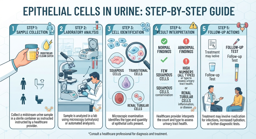
Because contamination is the number one cause of a worrying result, the most useful thing you can do is collect a proper clean-catch midstream sample. Here is the exact process I give my patients:
- Wash your hands with soap and water.
- Cleanse the genital area. Men: retract the foreskin if present and wipe the glans with the provided towelette. Women: separate the labia and wipe front to back.
- Begin urinating into the toilet for the first few seconds. This flushes cells and bacteria from the distal urethra.
- Move the sterile cup into the stream and collect roughly 30 to 60 mL without touching the inside of the cup.
- Finish urinating into the toilet.
- Cap the container and return it promptly. Samples sitting at room temperature for more than about two hours can alter cell counts and encourage bacterial growth.
- Tell the lab or clinician about any recent catheterization, sexual activity, menstruation, or antibiotics.
In women with persistent contamination, or whenever a clean specimen is essential, a catheterized specimen may be the better option.
Epithelial Cells in Urine: Use Cases and Applications in Clinical Practice
Urinary epithelial cells are not just an incidental finding. Clinicians use them in several ways:
- Specimen quality control: High squamous counts tell us whether a urine culture result can be trusted.
- Infection workup: Together with white blood cells, nitrite, and leukocyte esterase, they help differentiate cystitis from contamination.
- Kidney injury assessment: RTECs and granular casts help distinguish tubular from prerenal causes of acute kidney injury.
- Transplant monitoring: Increased tubular cells can prompt further evaluation for rejection or drug toxicity.
- Cancer evaluation: Atypical urothelial cells prompt urine cytology, cystoscopy, and imaging.
- Andrology workup: In men with urethral symptoms, epithelial findings guide testing for urethritis and prostatitis.
Why This Finding Is Often Misunderstood
Three patterns lead to unnecessary anxiety or missed diagnoses:
- Treating the number instead of the context. A patient with high squamous cells and no symptoms often receives antibiotics that are not needed.
- Assuming every epithelial cell is “just contamination.” RTECs and atypical urothelial cells are not contamination and can be overlooked when a report simply says “epithelial cells present.”
- Ignoring recurrent findings. One sample proves little. Repeated abnormal samples in a man over 35 who smokes, for example, deserve a proper evaluation.
Clinical Note: How This Affects Daily Life
Patients rarely tell you this at the first visit, but an “abnormal urine test” can quietly dominate a person’s life. I see men who stop drinking fluids before meetings to avoid bathroom trips, which concentrates their urine and worsens irritation. Others lose sleep from repeated nighttime checks of urine color, or avoid intimacy for fear of an undiagnosed infection. Health anxiety around a lab result is a real clinical issue. A clear explanation, and a clear plan, is often the first effective treatment.
A Real Patient’s Journey
The patient: A 46-year-old man, smoker for 20 years, who came to me after three urinalyses over eight months, each reporting “moderate epithelial cells.”
What he had been told: His primary care visits led to two courses of antibiotics for presumed UTI, and a third clinician reassured him it was “just contamination.” His symptoms (mild urgency, occasional weak stream) never fully resolved, and he stopped asking questions.
What I found: On careful review, the lab report separated cell types: squamous cells were low, but transitional cells were consistently elevated, and microscopic hematuria (3 to 5 red blood cells per HPF) had appeared on two samples. Urine culture was negative each time. Given his age, smoking history, and persistent microscopic hematuria, he met criteria for a formal hematuria evaluation under the AUA/SUFU guideline.
The workup: Cystoscopy, upper tract imaging (CT urography), and urine cytology. Cystoscopy revealed a small papillary lesion. Pathology confirmed a low-grade, non-muscle-invasive bladder tumor, resected transurethrally (TURBT).
The outcome: Because it was caught early, he needed no major surgery. He now has scheduled surveillance cystoscopy, has quit smoking, and his urinary symptoms improved after treatment of coexisting BPH.
The lesson: Most of the time, epithelial cells in urine mean nothing serious. But persistent findings, the wrong cell type, or blood in the sample should never be dismissed because earlier results “looked like contamination.”
Treatment Options: Treating the Cause, Not the Cell
There is no treatment for epithelial cells themselves. We treat what is producing them.
| Cause | Typical approach |
| Contamination | Repeat a proper clean-catch (or catheterized) sample; no medication needed |
| Bacterial cystitis | Targeted antibiotics based on culture and sensitivity |
| Urethritis (e.g., chlamydia, gonorrhea) | Pathogen-specific antibiotics; treat partners; abstain until treatment is complete |
| Kidney stones | Hydration, pain control, medical expulsive therapy, or ureteroscopy/lithotripsy depending on size and location |
| BPH or prostatitis | Alpha-blockers, 5-alpha-reductase inhibitors, or antibiotics where bacterial prostatitis is confirmed |
| Tubular injury (RTECs) | Nephrology evaluation; stop offending drugs; manage volume status and underlying disease |
| Urothelial lesion | Cystoscopy with TURBT, followed by surveillance and risk-adapted therapy |
Cystoscopy vs. Urine Cytology: Which Do I Prefer?
Patients often ask which test they “really” need if atypical cells are suspected.
- Urine cytology is non-invasive and good at detecting high-grade tumors, but it can miss low-grade lesions.
- Cystoscopy lets me see the bladder lining directly and take biopsies, and it remains the reference standard for bladder evaluation.
My preference: In a patient with hematuria and risk factors (age over 40, smoking, occupational exposure), I favor cystoscopy first, with cytology as an adjunct. In a younger, low-risk patient with no blood and a clearly contaminated sample, I would simply repeat the urinalysis first. The decision depends on age, smoking history, degree of hematuria, and persistence, which is exactly the risk-stratified approach the AUA guideline encourages
Recovery Timeline: What to Expect
- Contamination: A repeat sample is typically normal immediately.
- Uncomplicated UTI: Symptoms commonly improve within 24 to 72 hours of appropriate antibiotics; urinalysis typically normalizes within one to two weeks.
- Urethritis: Symptoms usually ease within days; retest per clinician guidance.
- Kidney stones: Small stones may pass within days to a few weeks; larger stones may need a procedure.
- Tubular injury: Recovery depends on the cause and can range from days to months; follow-up creatinine and urinalysis are essential.
- Bladder lesion after TURBT: Recovery is generally days to a couple of weeks, with surveillance cystoscopy at intervals set by your urologist.
When to Seek Immediate Medical Attention
Do not wait for a routine appointment if you have:
- Fever with chills and flank pain
- Visible blood or clots in your urine
- Inability to urinate
- Severe, sudden pain in the side, groin, or testicle
- Markedly reduced urine output or sudden swelling
- Confusion or lightheadedness along with urinary symptoms
Frequently Asked Questions
References
- Simerville JA, Maxted WC, Pahira JJ. Urinalysis: a comprehensive review. Am Fam Physician. 2005;71(6):1153-1162. https://www.aafp.org/pubs/afp/issues/2005/0315/p1153.html
- Kouri T, Fogazzi G, Gant V, et al. European Urinalysis Guidelines. Scand J Clin Lab Invest Suppl. 2000;231:1-86.
- Barocas DA, Boorjian SA, Alvarez RD, et al. Microhematuria: AUA/SUFU Guideline. J Urol. 2020;204(4):778-786. https://www.auanet.org/guidelines-and-quality/guidelines/microhematuria
- Fogazzi GB, Garigali G. The clinical value of urine sediment examination. Clin Chem Lab Med. (see journal for edition-specific citation.)
- Wise GJ, Schlegel PN. Sterile pyuria. N Engl J Med. 2015;372(11):1048-1054.