As heart disease is one of the most common causes of death globally, accurate diagnosis and physician treatment play a critical role in minimizing heart-complications. Two commonly performed but indispensable diagnostic tests in relation to heart disease are cardiac catheterization and coronary angiography. While the terms are often used interchangeably, they are two separate procedures with distinct roles in detecting and treating problems with the heart.
Cardiac catheterization and coronary angiography are two invasive diagnostic and therapeutic modalities that allow direct access to the coronary level and assistants in the management of heart conditions particularly the coronary artery disease (CAD).We will delve into their purpose, techniques, risks and recovery, explaining why they matter in assessing and treating your heart health.
Faced with the deadly consequences associated with heart disease, medical practitioners must depend on accurate diagnostic techniques. These techniques give important information about the heart’s function and anatomy, allowing directed interventions. While these procedures differ, they share a similar purpose to uncover hidden cardiovascular issues so that patients can receive the most appropriate and effective care in order to preserve cardiac strength.
Understanding Cardiac Catheterization
Cardiac catheterization is an important minimally invasive diagnostic and therapeutic approach to a large number of heart diseases. It involves putting a thin, flexible tube called a catheter into a blood vessel. This is usually done in the groin, wrist, or arm.
The tube is then guided to the heart while doctors watch real-time X-ray images. This procedure allows cardiologists to visualize the heart’s chambers, valves and major blood vessels and measure pressures inside the heart, take blood samples and even make interventions to correct abnormalities.
Cardiac catheterization can supplement non-invasive tests by providing information about the heart’s structure and function that noninvasive tests cannot, which is part of their versatility. This is crucial for assessing the severity of heart disease, for designing treatment plans and even for delivering life-saving therapy.
Purpose of Cardiac Catheterization: A Comprehensive Exploration
One type of diagnostic test that doctors may recommend in order for patients to have is a cardiac catheterization, depending on the needs of the patient. Here’s a closer look, why this test is recommended :
Coronary Artery Disease (CAD):
Chest pain (angina) or even a heart attack can occur if plaque builds up in the coronary arteries, blocking blood flow to the heart muscle. Cardiac catheterization enables detailed visualization of these blockages and can identify their location, severity (percentage of narrowing), and effect on blood flow.
Fractional Flow Reserve (FFR) and Instantaneous Wave-Free Ratio (iFR) are used during cardiac catheterization as advanced techniques to evaluate whether coronary artery occlusions are functionally significant.
These tests gauges the gradient of pressure across a blockage to identify whether it is responsible for a significant decrease in blood flow. That data is important for determining whether to treat a blockage with angioplasty or stenting.
During catheterization IVUS (intravascular ultrasound) and Optical Coherence Tomography (OCT) are also used. Both options contain a slight difference: IVUS uses sound waves for the imaging and OCT uses light waves, providing high-resolution images. These methods can give detailed insight into the composition of plaque and to what degree the arteries have narrowed.
Heart Valve Disorders:
The valves in the heart ensure that blood flows in one direction only. When these valves narrow (stenosis) or leak (regurgitation), it strains the heart. Cardiac catheterization is useful for accurately quantifying valve function since both pressure gradients across the valves and regurgitant volume can be used.
This is key to determining the severity of any valve disease, and to know whether a valve should be repaired or replaced. The transcatheter aortic valve replacement, TAVR, is a catheter-based, minimally-invasive procedure to replace a diseased aortic valve under cardiac catheterization settings.
Heart Muscle Performance:
Conditions like cardiomyopathy, heart failure and past heart attacks can weaken the heart muscle. Accurate determination of the heart’s ejection fraction (amount of blood ejected with each beat) and assessment of wall movement abnormalities regionally is done by cardiac catheterization.
It may also be used to identify portions of the heart muscle that have been damaged by a heart attack or other conditions.
Congenital Heart Defects:
Some congenital structural heart defects can be diagnosed, and sometimes treated, in the cardiac catheterization lab. These defects include atrial septal defects (ASDs), ventricular septal defects (VSDs), and patent foramen ovale (PFOs).
To some extent, these defects can be closed in a minimally invasive way with specialized devices that are released over a catheter and do not need for open-heart surgery.
Blood Pressure Measurement:
Cardiac catheterization also allows for direct measurement of pressures within the heart’s chambers and major blood vessels. This was particularly useful for diagnosing pulmonary hypertension, a condition whereby high blood pressure affects the arteries supplying blood to the lungs.
It can also help to diagnose other kinds of conditions affecting the pressures in the heart, such as constrictive pericarditis.
Treatment Procedures:
In addition to being a diagnostic procedure, cardiac catheterization enables intervention from a simple platform. These include:
Inflation of a balloon and placement of a stent to open blocked coronary arteries.
Balloon valvuloplasty to open narrowed heart valves.
References for the closure of congenital heart defects
Atherectomy, which cuts out plaque within the coronary arteries.
Myocardial biopsy, where a small sample of heart tissue is removed for analysis.
Understanding Coronary Angiography: Visualizing the Coronary Arteries
Coronary angiography is specifically a type of cardiac catheterization that allows good imaging of the coronary arteries, the blood vessels that supply oxygenated blood to the heart. During this process, a contrast dye that appears on X-ray is injected through the catheter, allowing Angiograms to be performed that provide detailed images of the coronary arteries.
These images, known as coronary angiograms, root out any blockages, narrowing or other issues that could get in the way of blood flow to the heart.
Coronary angiography allows for a detailed imaging of the coronary arteries and identification of obstructions that can cause myocardial ischemia or infarction. Stenosis severity and location of the narrowing in the arterial tree can be measured when contrast dye is injected into the arterial system and X-ray movies are taken by physicians.
Purpose of Coronary Angiography: Mapping the Heart’s Blood Supply
There are several critical reasons why Doctors recommend coronary angiography. Some of the reasons include:
Identifying Blockages in Coronary Arteries:
This procedure is the gold standard for detecting and characterizing blockages of the coronary arteries. The MAP depicts the coronary arteries in great detail: It shows where there is narrowing and how much there is of it.
That information is crucial for determining the extent of coronary artery disease and the development of an appropriate treatment strategy (medical therapy vs. angioplasty and stenting vs. coronary artery bypass grafting (CABG).
Blood Flow Measurement to the Heart:
Coronary angiograms visualize how well blood is flowing through the coronary arteries to the heart muscle.
It is important for assessing the functional significance of coronary artery blockages and predicting the risk for future cardiac events.
Evaluation of Chest Pain/Angina:
People in whom coronary artery disease is suspected, coronary angiography can provide a definitive diagnosis when such patients present with chest pain or other symptoms suggestive of angina.
This helps identify whether a patient’s chest pain is cardiac or noncardiac in nature so we can offer them the right treatment.
Post-Treatment Assessment:
Coronary angiography is commonly performed to visualize restoring blood supply to the heart muscle post angioplasty, stenting or CABG.
This can also aid in the detection of any complications or residual obstructions that might necessitate further treatment.
Analyzing Abnormal Results of Stress Tests:
Coronary angiography can result in further instructive diagnosis where the patient has an atypical stress test that is indicative of the underlying coronary artery disease.
This can identify the features explaining the silent stress test abnormality, and potentially direct future treatment.

Key Differences Between Cardiac Catheterization and Coronary Angiography
Although closely related, cardiac catheterization and coronary angiography have distinct purposes and applications. Understanding these differences is essential for patients and healthcare providers alike.
| Aspect | Cardiac Catheterization | Coronary Angiography |
| Purpose | Diagnoses and treats many heart conditions including CAD, valve disease, heart failure and congenital heart defects. | Uses imaging specifically to view the coronary arteries to diagnose and evaluate coronary artery disease. |
| Procedure | Includes a wide range of diagnostic and interventional techniques (pressure measurements, blood sampling, imaging, and even therapy). | Mainly requires injecting contrast dye into the coronary arteries and taking X-ray image. |
| Treatment Capability | Includes interventional procedures like angioplasty, stenting, valve repair, and closure of congenital defects. | Therapeutic interventions are not performed during coronary angiography, this is strictly a diagnostic procedure. |
| Scope | Wide range, including the entire heart and great blood vessels. | Heart and coronary arteries only |
Detailed Step-by-Step Procedure: Navigating the Process
While cardiac catheterization and coronary angiography are performed in a similar manner and require similar preparation, their diagnostic goals differ widely. Then, let’s go through the full steps of these procedures:
Step 1: Preparation: Ensuring Patient Safety and Comfort
Pre-Procedure Fasting
Typically there is direction not to eat 6 to 8 hours before a procedure. Preferably to lower the risk of vomiting and nausea that can occur during and after the process, particularly if ordered contrast dye or sedatives.
Additionally, this prolonged period of fasting ensures that the stomach is free of any contents, minimizing the risk of aspiration in the event of any potential complications with the patient.
Blood Tests
Complete blood tests are performed to check kidney function, clotting ability, and electrolyte balance.
Kidney function is especially crucial, because the contrast dye used in both is cleared through the kidneys. Patients with chronic kidney disease may be at greater risk for contrast-induced nephropathy (CIN), wherein contrast dye harms the kidneys.
A crucial test is the Glomerular Filtration Rate (GFR). The patient is being tested for clotting studies like prothrombin time (PT), and partial thromboplastin time (PTT). If the patient is on blood-thinning medications, then it may be necessary to down-dose them prior to the intervention.
Electrolytes are checked to make sure that the patient does not have a heart rhythm problem.
Medication Review
Extensive review of the patient’s current medications is necessary. Some meds, like metformin (for diabetes), may be temporarily paused prior to the procedure, as they can have interactions with the contrast dye.
Blood thinning medicines are a significant concern to treat.
Informed Consent
The procedure is explained to the patient extensively, including its risks, benefits, and alternatives. They are allowed to ask questions and give informed consent.
Sedation
Usually, sedatives are given to the patient to ensure that they are calm and not anxious in the entire process. The patient is awake and can follow commands.
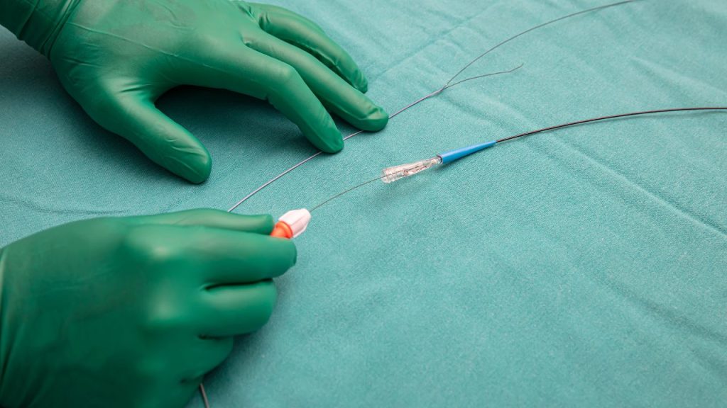
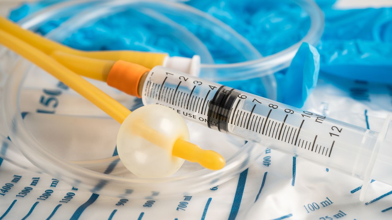
Step 2: Inserting the Catheter: Accessing the Heart
Puncture Site Preparation
The site where the needle will puncture the skin (usually the radial artery in the wrist, femoral artery in the groin or brachial artery in the arm) is cleaned and sterilized.
A local anesthetic is injected to numb the area, decreasing the pain during catheter insertion. As the radial artery is associated with fewer complications, it is gaining popularity for this procedure.
Catheter Insertion
A needle is passed into the selected artery, and it is then passed over with the thin wire.
The needle is taken out, and sustains (a short, empty tube) is put over the wire.
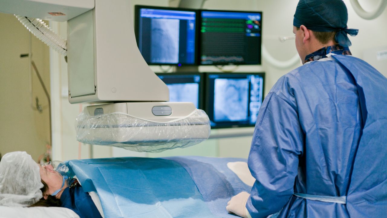
The wire is pulled out and the catheter itself can be carefully navigated through the sheath and into the heart using real-time X-ray imaging (fluoroscopy).
Guiding the Catheter
Fluoroscopy helps the cardiologist see where the catheter is being guided towards the heart or specific arteries.
Step 3: Imaging and Diagnosis: Unveiling the Heart’s Secrets
Cardiac Catheterization:
Diagnostic measurements can be taken once the catheter is positioned.
In this procedure, the heart chambers and large blood vessels are measured for pressure to evaluate heart function as well as for any abnormalities.
Blood tests may be performed to determine oxygen levels and heart function.
You can inject contrast dye into the left ventricle. This helps perform ventricular angiography. It is used to assess the heart’s pumping ability.
Biopsies can be taken.
Coronary Angiography:
The same smaller catheter is then advanced into the coronary arteries for coronary angiography.
A healthcare professional injects the contrast dye through the catheter. Then, the medical team takes several X-ray pictures, called coronary angiograms.
These images show the location and extent of any blockage or narrowing in the coronary arteries.
Multiple views are taken.
Step 4: Post-Procedure Care: Ensuring a Smooth Recovery
Catheter Removal:
Once completed, a catheter is removed with caution.
A person presses the puncture site for several minutes to stop the bleeding.
A healthcare provider may use a closure device at the puncture site if they accessed the femoral artery.
Monitoring
The patient is watched for a few hours after the procedure. This is to make sure their heart rhythm, blood pressure, and puncture site stay stable.
ECGs (Electrocardiograms) done to study heart rhythm.
Medical staff monitor the puncture site for bleeding or hematoma formation.
Recovery
Patients are generally advised to rest for a few hours afterward.
If a healthcare provider accesses the femoral artery, patients may need to lie flat for a few hours. This helps the puncture site to clot.
If doctors use the radial artery, recovery is faster, and patients can typically return to their normal activities sooner.
Discharge Instructions
The patients get written instructions about how to care for the puncture site. They also learn which medications to take and when to see their cardiologist again.
They also receive guidance on lifestyle changes, including a heart-healthy diet, exercise and stopping smoking.

Risks and Potential Complications: What Can Go Wrong
Cardiac catheterization and coronary angiography are generally safe procedures, carrying a small but significant risk of complication. Some of the risks include:
Bleeding or Hematoma
A common complication is bleeding or hematoma (an accumulation of blood out of blood vessel) in the puncture site.
Infection
Infection at the puncture site is a rare but known complication.
An allergic reaction to contrast dye
Patients can have a mild skin rash or severe allergic reaction to the dye called called anaphylaxis.
Kidney Injury (Contrast Induced Nephropathy)
The contrast dye is harmful to the kidneys, especially patients who have existing kidney disease.
Arrhythmias
Irregular heart rhythms (arrhythmias) may happen during the procedure.
Recovery and Aftercare: Supporting Healing and Heart Health
The need for a good recovery and aftercare plan are important here to healing and heart health.
Immediate Recovery
After the procedure, patients are monitored closely for a few hours.
The medical staff monitors bleeding or hematoma at the puncture site.
Doctors advise patients to drink lots of fluid to help clear the contrast dye from their kidneys.
Ongoing Care
Patients are instructed to eliminate vigorous activity for several days following the procedure.
The instructor teaches them how to care for the puncture site and watch for signs of infection.
Doctors may also prescribe blood thinners or antiplatelet medications to help prevent blood clots.
A follow up with the cardiologist is essential.
Lifestyle Modifications
Heart health starts with a heart-healthy lifestyle, which is important for managing your heart health in the long run. This includes:
Diet
A well-balanced diet that includes fruits and vegetables, whole grains, and lean protein.
Reducing saturated and trans fats, cholesterol and sodium.
Experts frequently suggest the DASH (Dietary Approaches to Stop Hypertension) diet or the Mediterranean diet.
Smoking Cessation
The best thing patients can do to improve their heart health is to quit smoking.
Stress Management
Some techniques are yoga, meditation or deep breathing exercises to release stress.
Weight Management
Keeping a healthy weight is extremely important.
Comparing Benefits and Limitations: A Balanced Perspective
Both cardiac catheterization and coronary angiography have their advantages and limitations in the diagnosis and management of heart diseases.
Benefits:
Accurate Diagnosis
These tests yield exceptionally precise information regarding the anatomy and physiology of the heart and coronary vessels.
They can see blockages, valve problems and other abnormalities that other (non-invasive) tests may not detect.
Treatment Guidance
The data from these procedures is important for cardiologists. They use it to decide the best treatment plan, like medical therapy, angioplasty, stenting, or surgery.
Immediate Intervention
Cardiac catheterization also enables immediate treatment, such as angioplasty or stenting, if a major blockage is identified.
Limitations:
Invasive Nature
These procedures are invasive and have a small risk of complications, including bleeding, infection and allergic reactions.
Contrast Dye Risks
The contrast dye used in these procedures can harm the kidneys. This is especially true for people with kidney disease.
Invasive Diagnostics (Coronary Angiography)
Coronary angiography is a diagnostic procedure and cannot treat blockages.
Radiation Exposure
The fluoroscopy that guides the catheter exposes the patient to radiation.
Conclusion: Empowering Patients with Knowledge
Cardiac catheterization and coronary angiography are critical components of diagnosis and management of heart disease. Cardiac catheterization allows for multiple diagnostic and therapeutic interventions, while coronary angiography allows visualization of coronary anatomy.
With knowledge about the reasons, methods, risks, and benefits of these tests, patients can take an active role in their health. They can work with their cardiologist to achieve the best heart health. Early detection and intervention is the key which prevents serious cardiac events and improve quality of life.
No one can tell you what is best for your health. Your situation is unique, so medical advice is very important.